| Line of Defense | Timeline | Cells | Antigen Dependency | Examples |
Innate | First | Immediate response (0 -96 hours) | Natural killer cells, macrophages, neutrophils, dendritic cells, mast cells, basophils, eosinophils | Independent | Skin, hair, cough, mucous membranes, phagocytes, granulocytes |
Adaptive | Second | Long term (>96 hours) | T and B lymphocytes | Dependent | Pus, swelling, redness, pain, T and B lymphocyte response |
 |
| Figure 1. DCs—Messengers between Innate and Adaptive Immune Systems |
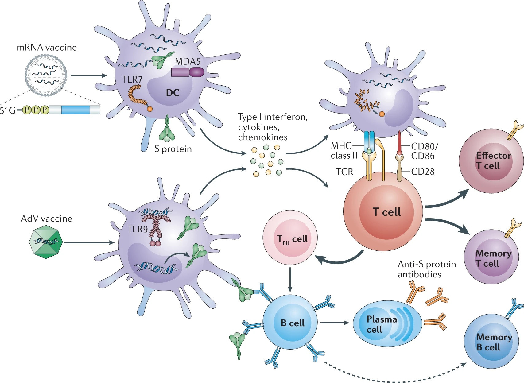 | ||
| Figure 2. COVID-19 vaccines immune activation modes (Source: [8]) |
Dendrite Cells
Dendritic cells (DC) are among the first cells to encounter pathogens/damage in peripheral tissues and, upon activation, DCs migrate to lymph nodes where they activate and educate T cells to initiate the immune response. DCs present pathogen-derived antigen to T cells and drive T cell differentiation into particular effector cells through the expression and secretion of co-stimulatory molecules and cytokines respectively.
Dendritic cells (DCs), named for their probing, ‘tree-like’ or dendritic shapes, are responsible for the initiation of adaptive immune responses and hence function as the ‘sentinels’ of the immune system.
During pathogen invasion, resident iDCs (DCs in immature state) detect intruders via pattern recognition receptor (e.g. TLRs) capture antigens and quickly leave the tissue. They crawl through the cells, cross the endothelium of lymphatic vessels and migrate to the draining lymph nodes (LN) in response to a number of chemokines such as CCL19 and CCL21.
During their migration from the peripheral tissues, DCs undergo phenotypical and functional maturation. Most remarkably, they stop capturing antigens while up-regulating the expression of co-stimulatory molecules such as CD80 and CD86 and the chemokine receptor CCR7, and secrete pro-inflammatory cytokines such as TNF-α and IL-12. After reaching the subcapsular sinus of the LN, DCs move to T-cell zones. Here, the interdigitating DCs are actively involved in the presentation of antigens to T cells.
- Professional antigen-presenting cells
- They are abundant at body surfaces and within tissues, where they sense and sample the environment for self- and non–self-antigens.
- Mononuclear phagocyte system (MPS)
- MPS comprises DCs, monocytes and macrophages
- 3 major subsets of DCs
- Plasmacytoid DCs, Conventional DCs, and Monocyte-derived DCs—are characterized by distinct origins, receptors, and functions.
- Undergoing a process of “maturation” upon antigen capture
- Which are exemplified by:
- Enhanced antigen processing
- Induction of major histocompatibility complex (MHC) molecules, co-stimulatory molecules (CD80/86), and cytokine production.
- Migrating to primary and secondary lymphoid organs
- Where they present processed antigens to naïve T cells to induce immunity or tolerance.
 |
| Figure 3. Schematic representation of the host immune response against microbial pathogens (Source: [1]). |
Functional Specialization of T Helper cells
Abs are complemented by T cells responding specifically to viral peptides presented by MHC class I and II molecules. CD8+ T cells are famous for their ability to lyse virus-infected cells expressing class I molecules complexed with viral peptides, but CD4+ T cells can also kill cells that express MHC class II molecules presenting viral peptides.[11]
Unlike class I molecules, which are constitutively expressed on nearly all cell types, class II molecules are expressed by immune cells and a few non-immune cells (including type II pneumocytes, a target cell for many respiratory viruses), though they are induced on many cell types by interferons. CD4+ T cells also play an essential role in B cell Ig class switching, somatic mutation, and memory cell formation.
These cells differentiate from naive T cells in response to signals from antigen presenting cells during activation and local microenvironmental cues.
The functional specialization of TH cells is conferred by the expression of T cell subset-specific transcription factors (TFs) that coordinate genetic programs to direct production of signature cytokines and surface molecules mediating interactions with other cells.[4]
- Th1 cells
- Express the TF T-bet and the cytokine interferon (IFN)-γ and mediate responses to intracellular pathogens
- Th2 cells
- Th17 cells
- Synthesize RORγt and IL-17 and limit extracellular bacteria and fungi, particularly at mucosal surfaces[5]
- Regulatory T cells (Tregs)
- Express the TF Foxp3[6]and modulates immunity by dampening effector T cell activation and proliferation
- T follicular helper cells (Tfh)
- Express Bcl-6 and a number of cell surface markers including CXCR5, PD1 and ICOS
- Play a critical role in protective immunity helping B cells produce antibody against foreign pathogens
References
- Belkaid, C. A. Piccirillo, S. Mendez, E. M. Shevach, D. L. Sacks, Nature 420, 502 (2002)
- T follicular helper cells (British Society for Immunology)
- The Innate vs. Adaptive Immune Response
- Polyamine metabolism is a central determinant of helper T cell lineage fidelity
- Y. Kanno, G. Vahedi, K. Hirahara, K. Singleton, J.J. O’Shea. Transcriptional and epigenetic control of T helper cell specification: molecular mechanisms underlying commitment and plasticity. Annu. Rev. Immunol., 30 (2012), pp. 707-731
- J.D. Fontenot, M.A. Gavin, A.Y. Rudensky. Foxp3 programs the development and function of CD4+CD25+ regulatory T cells. Nat. Immunol., 4 (2003), pp. 330-336.
- Dendritic Cell Control of Immune Responses
- COVID-19 vaccines: modes of immune activation and future challenges
- Designing spatial and temporal control of vaccine responses (good)
- Effect of Natural Compounds on NK Cell Activation
- NK cell-activating compounds: vitamins belonging to classes A, B, C, D, and E, polysaccharides, lectins, and a number of phytochemicals
- Antigenic drift: Understanding COVID-19 (good)
No comments:
Post a Comment
Note: Only a member of this blog may post a comment.