Sunday, November 21, 2021

Health Benefits of Selenium

Selenium (Se) is an essential trace element (for example, you only need to eat 1-2 Brazil Nuts per day; see Table 2) of high importance for human health . Studies have already identified associations between selenium deficiency and increased morbidity and mortality from viral infections, cardiovascular, and thyroid diseases, as well as prostate, gastrointestinal, and breast cancers. But, you do need to avoid overdosing on Se as warned by CDC:[17]
Brazil nuts contain very high amounts of selenium (68–91 mcg per nut) and could cause selenium toxicity if consumed regularly. Acute selenium toxicity has resulted from the ingestion of misformulated over-the-counter products containing very large amounts of selenium. In 2008, for example, 201 people experienced severe adverse reactions from taking a liquid dietary supplement containing 200 times the labeled amount. Acute selenium toxicity can cause severe gastrointestinal and neurological symptoms, acute respiratory distress syndrome, myocardial infarction, hair loss, muscle tenderness, tremors, lightheadedness, facial flushing, kidney failure, cardiac failure, and, in rare cases, death.


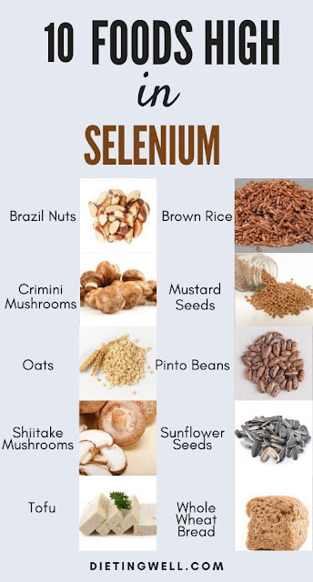
Figure 1.  10 foods high in Selenium (Source: dietingwell.com)


Figure 2.  Selenium absorption, metabolism, and distribution (Source: [12])

Absorption/ Metabolism / Distribution


Selenium is found in foods and nutritional supplements in 2 forms
After intestinal absorption, selenium forms are converted into hydrogen selenide (H2Se), a metabolic intermediate incorporated into selenoproteins, in the form of selenocysteine


Figure 3.  Selenium supplementation boosts TFH cells in mice and humans (Source: [7])

Important Roles of Selenoproteins


Se performs its main functions in the form of selenoproteins.  A wide range of these selenoproteins are linked to redox signaling, oxidative burst, calcium flux, and the subsequent effector functions of immune cells being grouped into families such as:
In addition, the Selenoprotein P (or SELENOP) acts as the main selenium transporter for peripheral tissues also performing extracellular antioxidant function (see Figure 2). 

Thus, selenium plays a role in antioxidant, anticarcinogenic, anti-inflammatory, redox and immune-cell function as well as in the regulation of thyroid hormone metabolism:
  • Anti-inflammatory
    • Findings suggest that UV damage to the epidermis affects deeper layers of the skin and even blood and other tissues
      • Diets enriched with antioxidant nutrients, including selenium, beta-carotene, vitamin E, and vitamin C, inhibit the formation of UV-induced tumors.[4]
    • A major role of the exogenous antioxidants, vitamins C and E (a-tocopherol), b-carotene, and selenium, is to act as efficient scavengers of reactive oxygen radicals, thereby protecting against oxidative damage.[14]
  • Antiviral
    • Se/selenoproteins are relevant in the viral pathogenicity, notably reducing proliferation of T cells, lymphocyte-mediated toxicity and NK cell activity, all of which are crucial for antiviral immunity. 
    • Selenoproteins partly reduce oxidative stress generated by viral pathogens.
    • The available studies support the belief that selenium may be of relevance in the infection with SARS-CoV-2 and disease course of COVID-19.
      • There is a mechanism proposed by Zhang et al.[10] by which selenium might suppress the life cycle and mutation to virulence of SARS-COV-2 while attenuating viral-induced oxidative stress, organ damage and the cytokine storm.
  • Redox
    • Selenoproteins also regulate or are regulated by cellular redox tone, which is a crucial modulator of immune cell signaling. 
      • The cellular redox environment is a balance between the production of reactive oxygen species (ROS), reactive nitrogen species (RNS), and their removal by antioxidant enzymes and small-molecular-weight antioxidants.
    • Redox-active selenium metabolites are involved in the anti-viral action of selenium in mice and humans.[15]
  • Immune-cell activity
    • Se is useful for the competency of the cellular component of both innate and adaptive immunity.
    • Inhibition of ferroptosis (i.e., a form of regulated cell death) via selenium supplementation promotes the survival of follicular helper T cells (TFH), boosting the germinal center and antibody response following vaccination in mice and people (see Figure 3).[7]
    • On a cellular level, Se status may influence various leukocytic functions including adherence, migration, phagocytosis, and cytokine secretion. 
  • Regulation of thyroid hormone metabolism
    • Selenium in iodothyronine deiodinase, as selenocysteine, plays a crucial role in determining the free circulating levels of T3. Selenium deficiency can have implications in fall of T3 levels.
      • In target tissues, T4, the most abundant circulating thyroid hormone, can be converted to T3 by selenium-containing enzymes known as deiodinases.[11]
Table 1: Selenium Content of Selected Foods [16]
FoodMicrograms
(mcg) per
serving
Percent
DV*
Brazil nuts, 1 ounce (6–8 nuts)544989
Tuna, yellowfin, cooked, dry heat, 3 ounces92167
Halibut, cooked, dry heat, 3 ounces4785
Sardines, canned in oil, drained solids with bone, 3 ounces4582
Ham, roasted, 3 ounces4276
Shrimp, canned, 3 ounces4073
Macaroni, enriched, cooked, 1 cup3767
Beef steak, bottom round, roasted, 3 ounces3360
Turkey, boneless, roasted, 3 ounces3156
Beef liver, pan fried, 3 ounces2851
Chicken, light meat, roasted, 3 ounces2240
Cottage cheese, 1% milkfat, 1 cup2036
Rice, brown, long-grain, cooked, 1 cup1935
Beef, ground, 25% fat, broiled, 3 ounces1833
Egg, hard-boiled, 1 large1527
Bread, whole-wheat, 1 slice1324
Baked beans, canned, plain or vegetarian, 1 cup1324
Oatmeal, regular and quick, unenriched, cooked with water, 1 cup1324
Milk, 1% fat, 1 cup815
Yogurt, plain, low fat, 1 cup815
Lentils, boiled, 1 cup611
Bread, white, 1 slice611
Spinach, frozen, boiled, ½ cup59
Spaghetti sauce, marinara, 1 cup47
Cashew nuts, dry roasted, 1 ounce35
Corn flakes, 1 cup24
Green peas, frozen, boiled, ½ cup12
Bananas, sliced, ½ cup12
Potato, baked, flesh and skin, 1 potato12
Peach, yellow, raw, 1 medium00
Carrots, raw, ½ cup00
Lettuce, iceberg, raw, 1 cup00

*DV = Daily Value.

References

  1. Health Benefits of Iodine (Travel and Health)
  2. Beck MA. Antioxidants and viral infections: host immune response and viral pathogenicity.  J Am Coll Nutr 2001; 20 (5 Suppl): 384S-388S, discussion 396S-397S.
  3. Garlic—a Vegetable, a Condiment, and a Medicine (Travel and Health)
  4. Health Benefits of Carotenoids (Travel and Health)
  5. What You Need to Know About Your Thyroid Health (Dr. Mercola)
  6. Selenium (Harvard School of Public Health; good)
  7. Selenium saves ferroptotic TFH cells to fortify the germinal center
  8. Nutritional risk of vitamin D, vitamin C, zinc, and selenium deficiency on risk andclinical outcomes of COVID-19: a narrative review
  9. Huang Z, Rose AH, Hoffmann PR. The role of selenium in inflammation and immunity: from molecular mechanisms to therapeutic opportunities. Antioxid Redox Signal 2012 Apr 1;16(7):705-43.
  10. Zhang J, Saad R, Taylor EW, Rayman MP. Selenium and selenoproteins in viral infection with potential relevance to COVID-19. Redox Biol 2020 Oct;37:101715
  11. Iodine (Linus Pauling Institute)
  12. Selenium in Human Health and Gut Microflora: Bioavailability of Selenocompounds and Relationship With Diseases
  13. The Role of Selenium in Inflammation and Immunity: From Molecular Mechanisms to Therapeutic Opportunities
  14. Vitamin C and the risk of developing inflammatory polyarthritis: prospective nested case-control study
  15. Yu L., Sun L., Nan Y., Zhu L.Y. Protection from H1N1 influenza virus infections in mice by supplementation with selenium: a comparison with selenium-deficient mice. Biol. Trace Elem. Res. 2011;141(1–3):254–261.
  16. U.S. Department of Agriculture, Agricultural Research Service. FoodData Central, 2019.
  17. Selenium—Fact Sheet for Health Professionals (NIH)
  18. Egg consumption improves vascular and gut microbiota function without increasing inflammatory, metabolic, and oxidative stress markers
Thursday, November 11, 2021

Aging—Knowing the Basics

Figure 1. The hallmarks of ageing (Source: [55])


The well-established nine hallmarks of aging include:[55]
  1. Genomic instability
  2. Shortening telomere length
  3. Epigenetic modifications
  4. Loss of proteostasis
  5. Deregulated nutrient sensing
  6. Mitochondrial dysfunction
    • SIRT3, an important stress-responsive deacetylase with cardio-protective and longevity enhancing properties involved in mitochondrial homeostasis, stem cells and tissue maintenance.[56,57]
  7. Cellular senescence
  8. Stem cell exhaustion
  9. Altered intracellular communication (Video 2)
have all been shown to be caused, at least in part, by sustained systemic inflammation.[45-54]

Video 1. Can ageing be delayed, stopped or even reversed? BBC News (YouTube link)

Inflammatory Clock (or iAge)


Inflammation plays a role in almost all chronic diseases of ageing including atherosclerosis, cancer, neurodegenerative diseases and diabetes.

From the blood immunome of 1,001 individuals aged 8–96 years, Sayed et al. developed a deep-learning method based on patterns of systemic age-related inflammation.[58] The resulting inflammatory clock of aging (iAge) tracked with multimorbidity, immunosenescence, frailty and cardiovascular aging, and is also associated with exceptional longevity in centenarians.[58]
In centenarians, iAge was on average, 40 years lower than their corresponding chronological age. Note that the lower your iAge is, the better.

iAge is demonstrated to be correlated with multi-morbidity and immunosenescence and can be used as a ‘metric’ for immunological health. Based on the results of iAge research, here are the findings with increasing iAge :
  • CXCL9 (an interferon-related chemokine)
    • CXCL9 is the most robust contributor to iAge
    • Aging endothelial cells express high levels of CXCL9, which induces mRNA down-regulation of the cardio-protective SIRT3 — a gene known to be important in aging and endothelial cell function.
    • CXCL9 was validated as an indicator of cardiovascular pathology independent of age
    • One root cause of CXCL9 overproduction is cellular aging per-se, which triggers metabolic dysfunction with production of damage-associated molecular patterns (DAMPs).[62]
  • B cell and T cell immune responses
    • A strong association with poor acute immune responses to cytokine stimuli was found, which is consistent with reports by two independent studies showing that high levels of baseline inflammatory markers correlate with weaker responses to hepatitis B and herpes zoster vaccine formulations.[59,60]
  • Potentiated monocyte responses
  • JAK-STAT response
    • Chronic inflammation is, at least in part, responsible for a reduced JAK-STAT response to cytokine stimulations in various leukocyte populations.[61]
This suggests that this immune ‘metric’ for human health versus disease may be useful as a companion diagnostic to inform physicians about patient’s inflammatory status, especially those with chronic diseases.
Their results indicate that CXCL9 and SIRT3 play an important role linking inflammation, cell metabolism, endothelial cell function and cardiovascular remodeling, which is consistent with prior work showing intricate interactions between inflammation and cell metabolism in tissue repair processes.[63]

Video 2. AGE Presents: Scott Leiser - Altered Cellular Communications (YouTube link)

Longevity Pathways


In Video 2 (must watch), Dr. Scott Leiser has shown that human may live longer by:
The common denominator of the above longevity pathways is stress—in the sense that either they are stresses directly or can turn on the stress response pathways indirectly (video 2 @7:52).  Note that under stress, organisms actually get healthier under low stress (hormetic effects). However, all of these longevity pathways also have some drawbacks or side effects in the real world (see Figure 3).

Figure 2.  Longevity pathway is conserved from yeast to humans (video 2 @6:30)

Figure 3.  Longevity pathways have drawbacks or side effects in the real world (video 2 @13:23)

Video 3. Living into your 90s (YouTube link)

Video 4.  Why We Age – And Why We Don't Have To (YouTube link)

Video 5.  Dr. David Sinclair: The Biology of Slowing & Reversing Aging  (YouTube link)

References

  1. SARS-CoV-2 causes senescence in human cells and exacerbates the senescence-associated secretory phenotype through TLR-3.
  2. REST/NRSF deficiency impairs autophagy and leads to cellular senescence in neurons.
  3. Nutrition and cellular senescence in obesity-related disorders.
  4. Deferoxamine accelerates endothelial progenitor cell senescence and compromises angiogenesis.
  5. Riboflavin transporter SLC52A1, a target of p53, suppresses cellular senescence by activating mitochondrial complex II.
  6. A cGAS-dependent response links DNA damage and senescence in alveolar epithelial cells: A potential drug target in IPF.
  7. The balance between NAD+ biosynthesis and consumption in ageing.
  8. RAS induced senescence of skin keratinocytes is mediated through Rho-associated protein kinase (ROCK).
  9. Radiation-induced liver injury and hepatocyte senescence.
  10. Oxidative stress in retinal pigment epithelium impairs stem cells: a vicious cycle in age-related macular degeneration.
  11. DNA methylation-based biomarkers of aging were slowed down in a two-year diet and physical activity intervention trial: the DAMA study.
  12. Virus-induced senescence is driver and therapeutic target in COVID-19.
  13. The hepatic senescence-associated secretory phenotype promotes hepatocarcinogenesis through Bcl3-dependent activation of macrophages.
  14. Necroptosis increases with age in the brain and contributes to age-related neuroinflammation.
  15. Cellular senescence promotes cancer metastasis by enhancing soluble E-cadherin production.
  16. Transcriptional features of biological age maintained in human cultured cardiac interstitial cells.
  17. H2S-mediated blockage of protein acetylation and oxidative stress attenuates lipid overload-induced cardiac senescence.
  18. Immunology of Aging: the Birth of Inflammaging.
  19. IL-1 Mediates Microbiome-Induced Inflamm-Ageing of Hematopoietic Stem Cells in Mice.
  20. Loss of polycomb repressive complex 1 activity and chromosomal instability drive uveal melanoma progression.
  21. Preclinical and clinical evidence of NAD+ precursors in health, disease, and ageing.
  22. Leaked Mitochondrial C1QBP Inhibits Activation of the DNA Sensor cGAS.
  23. S3QELs protect against diet-induced intestinal barrier dysfunction.
  24. Multi-omic profiling of primary mouse neutrophils predicts a pattern of sex and age-related functional regulation.
  25. Decreased proliferation of aged rat beta cells corresponds with enhanced expression of the cell cycle inhibitor p27KIP1.
  26. Exogenous melatonin prevents type 1 diabetes mellitus-induced bone loss, probably by inhibiting senescence.
  27. Astrocyte-Derived Extracellular Vesicles (ADEVs): Deciphering their Influences in Aging.
  28. Chronic inflammation-induced senescence impairs immunomodulatory properties of synovial fluid mesenchymal stem cells in rheumatoid arthritis.
  29. Molecular mechanisms of dietary restriction promoting health and longevity.
  30. Repetitive spikes of glucose and lipid induce senescence-like phenotypes of bone marrow stem cells through H3K27me3 demethylase-mediated epigenetic regulation.
  31. Protective effect of MitoQ on oxidative stress-mediated senescence of canine bone marrow mesenchymal stem cells via activation of the Nrf2/ARE pathway.
  32. Classical HDACs in the regulation of neuroinflammation.
  33. Environmental enrichment mitigates age-related metabolic decline and Lewis lung carcinoma growth in aged female mice.
  34. Effect of different exercise training intensities on age-related cardiac damage in male mice.
  35. A high fat, sugar, and salt Western diet induces motor-muscular and sensory dysfunctions and neurodegeneration in mice during aging: Ameliorative action of metformin.
  36. p38 MAPK-mediated loss of nuclear RNase III enzyme Drosha underlies amyloid beta-induced neuronal stress in Alzheimer's disease.
  37. Reversal of brain aging by targeting telomerase: A nutraceutical approach.
  38. Emerging roles of kisspeptin/galanin in age-related metabolic disease.
  39. Environmental enrichment modulates silent information regulator 1 (SIRT1) activity to attenuate central presbycusis in a rat model of normal aging.
  40. Lithium can mildly increase health during ageing but not lifespan in mice.
  41. The Evolution of the Hallmarks of Aging (good)
  42. N-acetyl cysteine ameliorates aortic fibrosis by promoting M2 macrophage polarization in aging mice.
  43. Environmental enrichment mitigates age-related metabolic decline and Lewis lung carcinoma growth in aged female mice (good)
  44. Effect of different exercise training intensities on age-related cardiac damage in male mice (good)
  45. Cavadas, C., Aveleira, C.A., Souza, G.F., and Velloso, L.A. (2016). The pathophysiology of defective proteostasis in the hypothalamus - from obesity to ageing. Nature reviews Endocrinology 12, 723–733.
  46. Efeyan, A., Comb, W.C., and Sabatini, D.M. (2015). Nutrient-sensing mechanisms and pathways. Nature 517, 302–310.
  47. Grivennikov, S.I., Greten, F.R., and Karin, M. (2010). Immunity, inflammation, and cancer. Cell 140, 883–899.
  48. Hunter, R.L., Dragicevic, N., Seifert, K., Choi, D.Y., Liu, M., Kim, H.C., Cass, W.A., Sullivan, P.G., and Bing, G. (2007). Inflammation induces mitochondrial dysfunction and dopaminergic neurodegeneration in the nigrostriatal system. Journal of neurochemistry 100, 1375–1386.
  49. Jurk, D., Wilson, C., Passos, J.F., Oakley, F., Correia-Melo, C., Greaves, L., Saretzki, G., Fox, C., Lawless, C., Anderson, R., et al. (2014). Chronic inflammation induces telomere dysfunction and accelerates ageing in mice. Nature communications 2, 4172.
  50. Lasry, A., and Ben-Neriah, Y. (2015). Senescence-associated inflammatory responses: aging and cancer perspectives. Trends in immunology 36, 217–228.
  51. Nathan, C., and Cunningham-Bussel, A. (2013). Beyond oxidative stress: an immunologist’s guide to reactive oxygen species. Nature reviews Immunology 13, 349–361.
  52. Oh, J., Lee, Y.D., and Wagers, A.J. (2014). Stem cell aging: mechanisms, regulators and therapeutic opportunities. Nature medicine 20, 870–880.
  53. Thevaranjan, N., Puchta, A., Schulz, C., Naidoo, A., Szamosi, J.C., Verschoor, C.P., Loukov, D., Schenck, L.P., Jury, J., Foley, K.P., et al. (2017). Age-Associated Microbial Dysbiosis Promotes Intestinal Permeability, Systemic Inflammation, and Macrophage Dysfunction. Cell host & microbe 21, 455–466 e454.
  54. Alpert, A., Pickman, Y., Leipold, M., Rosenberg-Hasson, Y., Ji, X., Gaujoux, R., Rabani, H., Starosvetsky, E., Kveler, K., Schaffert, S., et al. (2019). A clinically meaningful metric of immune age derived from high-dimensional longitudinal monitoring. Nature medicine 25, 487–495.
  55. The Hallmarks of Aging
  56. Bonkowski, M.S., and Sinclair, D.A. (2016). Slowing ageing by design: the rise of NAD+ and sirtuin-activating compounds. Nature reviews Molecular cell biology 17, 679–690.
  57. Lu, Y., Wang, Y.D., Wang, X.Y., Chen, H., Cai, Z.J., and Xiang, M.X. (2016). SIRT3 in cardiovascular diseases: Emerging roles and therapeutic implications. International journal of cardiology 220, 700–705.
  58. Sayed, N. et al. An inflammatory aging clock (iAge) based on deep learning tracks multimorbidity, immunosenescence, frailty and cardiovascular aging. Nature Aging (2021)
  59. Fourati, S., Cristescu, R., Loboda, A., Talla, A., Filali, A., Railkar, R., Schaeffer, A.K., Favre, D., Gagnon, D., Peretz, Y., et al. (2016). Pre-vaccination inflammation and B-cell signalling predict age-related hyporesponse to hepatitis B vaccination. Nature communications 7, 10369.
  60. Thevaranjan, N., Puchta, A., Schulz, C., Naidoo, A., Szamosi, J.C., Verschoor, C.P., Loukov, D., Schenck, L.P., Jury, J., Foley, K.P., et al. (2017). Age-Associated Microbial Dysbiosis Promotes Intestinal Permeability, Systemic Inflammation, and Macrophage Dysfunction. Cell host & microbe 21, 455–466 e454.
  61. Shen-Orr, S.S., Furman, D., Kidd, B.A., Hadad, F., Lovelace, P., Huang, Y.W., Rosenberg-Hasson, Y., Mackey, S., Grisar, F.A., Pickman, Y., et al. (2016). Defective Signaling in the JAK-STAT Pathway Tracks with Chronic Inflammation and Cardiovascular Risk in Aging Humans. Cell systems 3, 374–384 e374.
  62. Expression of specific inflammasome gene modules stratifies older individuals into two extreme clinical and immunological states
  63. Eming, S.A., Wynn, T.A., and Martin, P. (2017). Inflammation and metabolism in tissue repair and regeneration. Science 356, 1026–1030.
  64. Bruns DR, Drake JC, Biela LM, Peelor FF 3rd, Miller BF, Hamilton KL. Nrf2 signaling and the slowed aging phenotype: evidence from long-lived models. Oxid Med Cell Longev. 2015; 2015:732596.
  65. Hiebert P, Wietecha MS, Cangkrama M, Haertel E, Mavrogonatou E, Stumpe M, Steenbock H, Grossi S, Beer HD, Angel P, Brinckmann J, Kletsas D, Dengjel J, Werner S. Nrf2-mediated fibroblast reprogramming drives cellular senescence by targeting the matrisome. Dev Cell. 2018; 46:145–161.
    • This suggests that time-controlled activation of NRF2 may be critical for homeostasis in multicellular organism.
  66. A global clinical measure of fitness and frailty in elderly people
  67. DR. DAVID SINCLAIR: THE BIOLOGY OF SLOWING & REVERSING AGING
  68. New Insights into the Roles and Mechanisms of Spermidine in Aging and Age-Related Diseases

Sunday, November 7, 2021

The Link between TLR7 Variants and Critical COVID-19

Toll Like Receptor 7 (TLR7) is a Protein Coding gene. Diseases associated with TLR7 include:[3]
Among its related pathways are Viral mRNA Translation and Measles. Gene Ontology (GO) annotations related to this gene include transmembrane signaling receptor activity and double-stranded RNA binding (dsRBD). An important paralog of this gene is TLR8.

In this article, we will cover the below topics:
  • Genetic Variation
  • X-linked COVID-19 Risk Factor
  • Raw Data on Your Genetic Data (23andMe)
    • How Does 23andMe Report Genotypes?
    • All Markers Reported on 23andMe for TLR7 Gene

Genetic Variation


Genetic variation is the difference in DNA sequences between individuals within a population. Variants may be germline or somatic.

A variant in itself is not “pathogenic”, whether it can be causally related to a phenotype observed in a patient is determined by other factors. When a variant is described to "cause disease", the expert probably means “causes disease in a specific context”, e.g:

X-linked COVID-19 Risk Factor


Age and male sex are two prominent risk factors for developing life-threatening COVID-19 after SARS-CoV-2 infection. 

Asano et al. analyzed 1202 critical male COVID-19 patients to examine whether non-synonymous variants in genes on the X chromosome are a risk factor for developing COVID-19 pneumonia. TLR7 variants resulting in TLR7 deficiency occurred in 16 unrelated males, most of which were under age 60.[2]  
Plasmacytoid dendritic cells (pDCs), primary producers of type I interferon (IFN-I), from TLR7-deficient patients were unresponsive to TLR7 stimulation and displayed impaired production of IFN-I in response to SARS-CoV-2. These results identify X-linked recessive TLR7 deficiency as a genetic risk factor for COVID-19 pneumonia in males and demonstrate a key role for intact pDC IFN-I in protective immunity against SARS-CoV-2.
 

Immunodeficiency-74 (IMD74


Immunodeficiency-74 (IMD74) is also an X-linked recessive specific immunologic disorder characterized by the development of severe respiratory insufficiency in response to infection with the COVID19 coronavirus.[6] 

Affected individuals usually require mechanical ventilation in the ICU in order to survive. Laboratory studies show activation of the immune response and may show perturbation of some values, such as increased D-dimers and fibrinogen
In vitro functional studies of patient immune cells show impaired signaling through the TLR7 pathway, resulting in defective type I and type II interferon (IFN) responses. The patients reported to date did not have a history of immunodeficiency or chronic disease.[6]


Table 1.  TLR7 Genotype (all markers found for TLR7 Gene on 23andMe)

Raw Data on Your Genetic Data (23andMe)


23andMe uses genotyping, not sequencing, to analyze your DNA. Read [10] for more details.

How Does 23andMe Report Genotypes?


The 23andMe genotyping platform detects single nucleotide polymorphisms (SNPs).[10] A SNP is a DNA location, or "marker," in the genome that has been shown to vary among people in terms of the DNA base or bases. There are four DNA bases: 
So, for example, at the same genomic location, you might have a C and someone else might have a T. These DNA base differences are known as "variants."

For most SNPs on the 23andMe platform, the 23andMe Raw Data feature reports the marker name (usually an rsID or internal ID number), its exact genomic location, the possible variants at that marker (A, T, G, or C), and the specific variants you have, i.e. your genotype (See Table 1). 

Because you have two sets of autosomal chromosomes -- one from your mother and one from your father -- you usually have two variants at every location, and your genotype will be reported as a pair of variants, e.g. "G/A."

In some cases your genotype will be reported as a single variant because not all DNA is inherited in chromosome pairs e.g., mitochondrial DNA and, for the most part, the X and Y chromosomes in men).

Occasionally, for some SNPs on the 23andMe platform, your genotype may be reported as an insertion or deletion (--) of DNA bases instead of just a simple variant pair

Depending on the genomic location, either an insertion or deletion could represent the typical version of the SNP. In other words, there are some markers in which having an extra base (insertion) is the typical variant and having a deletion is the less common variant. Conversely, there are some places in the genome where an insertion is rare, making a deletion the typical variant at that location.

23andMe does not report on all possible insertions or deletions. In general, the ones reported on are small, spanning only one or a few bases.   


All Markers Reported on 23andMe for TLR7 Gene


On 23andMe, you can search for specific genes, markers, or positions of interest.  For example, here are the list of markers found by searching with TLR7 keyword:
Table 2.  TLR7 with NM_016562.4:c.3+4753C>T change

However, 23andMe uses genotyping, not sequencing, to analyze your DNA.  So, not all markers or positions are tested by 23andMe.  They only look for variants of interest at the time of testing.[10,11]
Genotyping can be performed through a variety of different methods, depending on the variants of interest and the resources available. For looking at many different variants at once, especially common variants, genotyping chips are an efficient and accurate method. They do, however, require prior identification of the variants of interest.